Receive Path of Typical Ultrasound System in Color Doppler Mode This application report looks at efficient implementation of the following typical color Doppler algorithms. Try these curated collections.
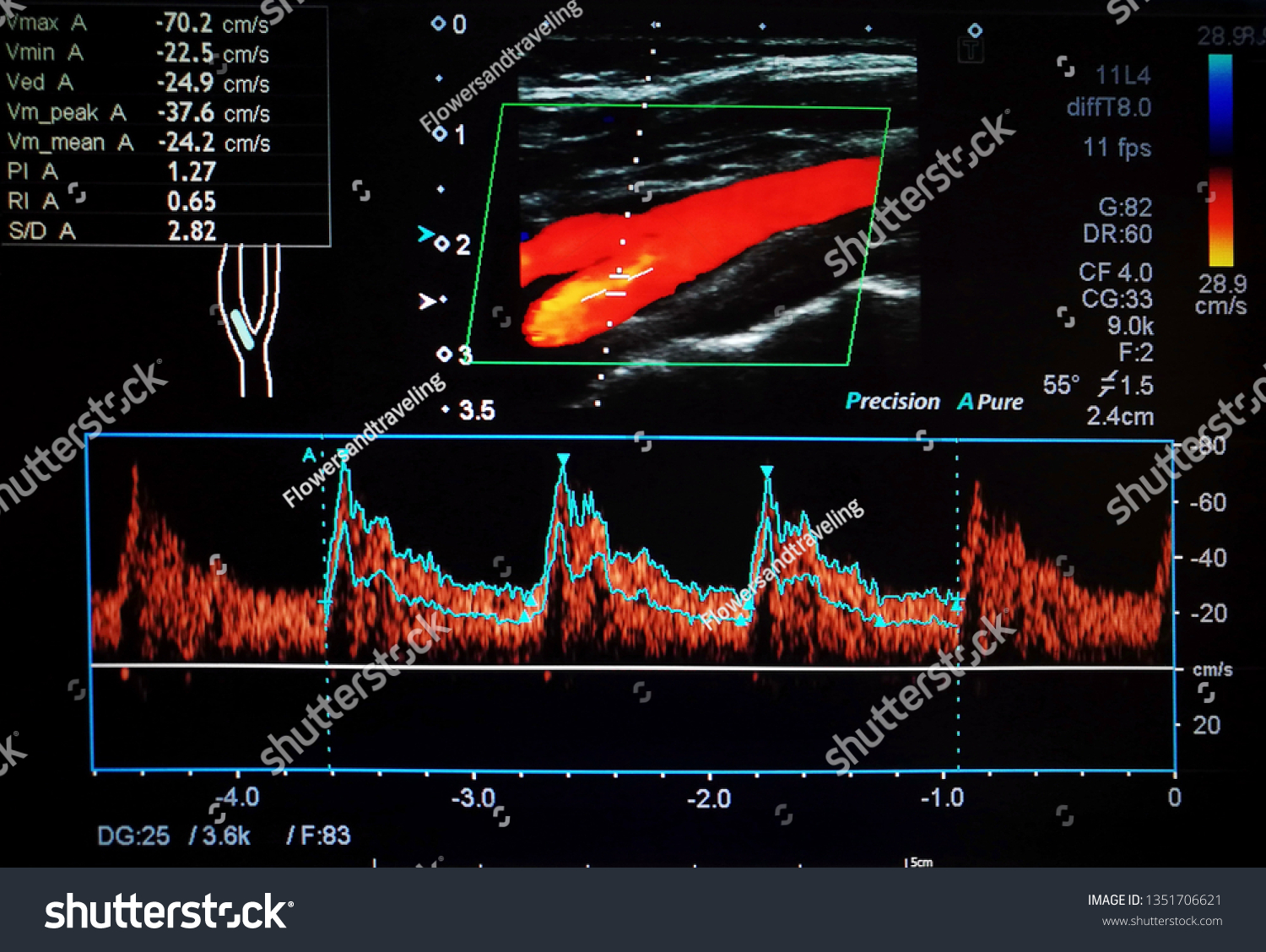
Color Doppler Ultrasound Imaging Normal Carotid Stock Photo Edit Now 1351706621
Flow that travels away from the transducer negative Doppler shift is depicted in blue and flow that is traveling toward the transducer positive Doppler shift is depicted in red with lighter shades of each color denoting higher velocities.

Color doppler ultrasound images. This test uses standard ultrasound to take images of blood vessels and organs. Transverse image through MPV 2D and color Doppler to show patency and direction. Power Doppler and Colour Doppler Ultrasound Power Doppler is a form that is more sensitive to slow flow but is described as one color eg.
Velocity is color-coded according to the color bar shown at the left side of the image red arrow. Such visualization is achieved by color-encoding Doppler information and displaying the colors as an overlay on the 2D image of the heart. The colors represent the speed and direction of blood flow within a certain area of the image color box.
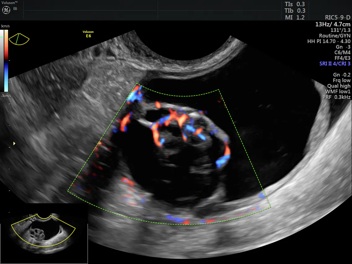
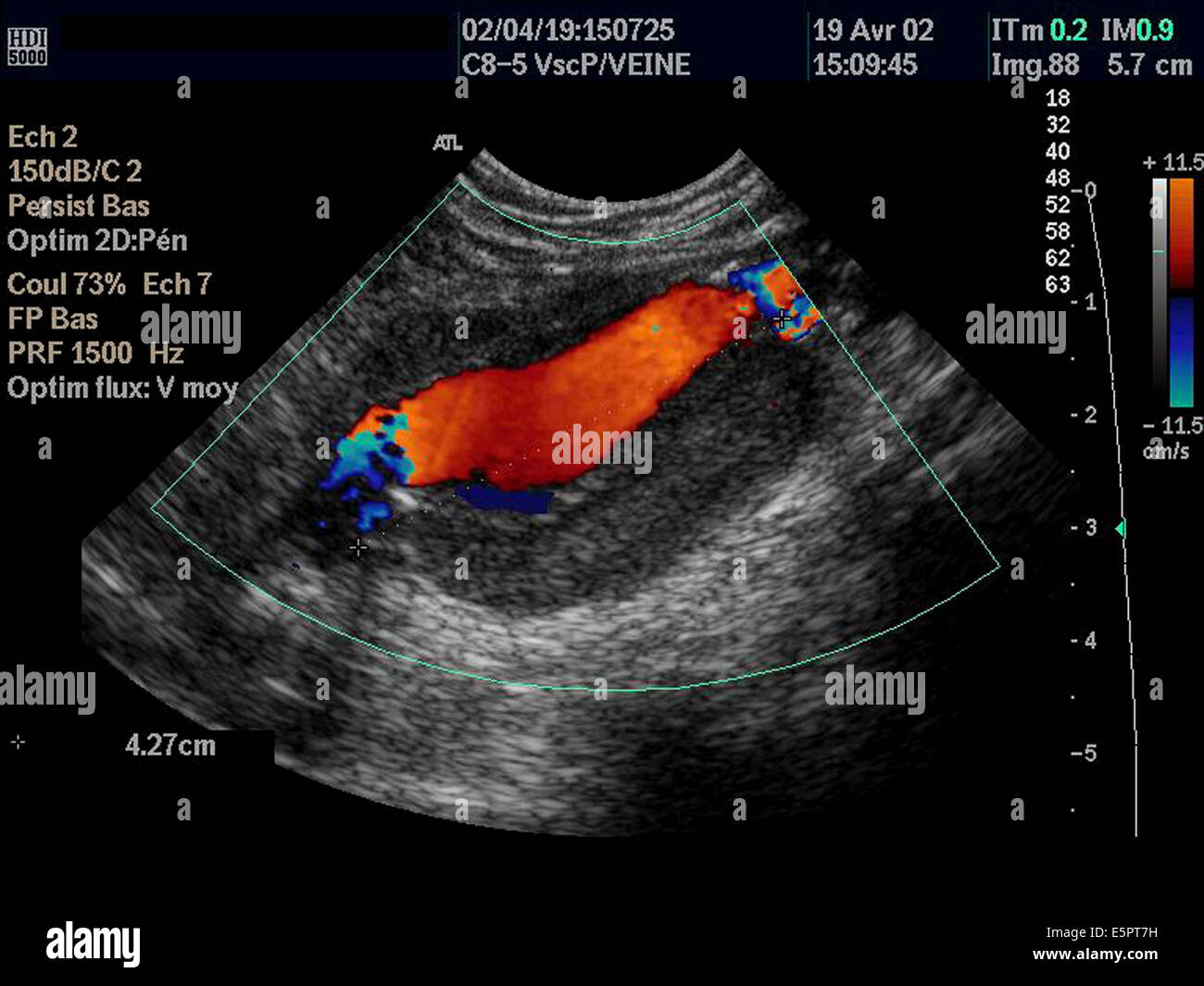
Color Doppler images are generally combined with grayscale images to display duplex ultrasonography images allowing for simultaneous visualization of the anatomy of the area. Then a computer turns the images into a graph as in spectral Doppler. The use of colour flow Doppler CFD or colour Doppler imaging CDI or simply colour Doppler sonography allows the visualisation of flow direction and velocity within a user defined areaA region of interest is defined by the sonographer and the Doppler shifts of returning ultrasound waves within are colour-coded based on average velocity and direction.
Among these parameters the color box color gain color velocity scale and inversion are frequently used during color Doppler US scanning. Color Doppler ultrasound image shows a grade 3 varicocele of the left side. See doppler ultrasound stock video clips.
The color box is a square area within the grayscale sonogram in which all color Doppler information is displayed Fig. One color is used to indicate flow toward the transducer and the other used to indicate flow away from the transducer. Uses color Doppler ultrasound images that span one heart cycle The consecutive set of frames used is called a cineloop.
Ultrasound doppler imaging is the ability to estimate and measure blood flow through various veins arteries and vessels. Curvilinear and phased array transducers have a radiating pattern of ultrasound beams that can produce complex color flow images depending on the orientation of the arteries and veins. Doppler ultrasound is unable to determine the specific location of velocities within the beam and cannot be used to produce color flow images.
Relatively inexpensive Doppler ultrasound systems are available which employ continuous wave probes to give Doppler output without the addition of B-mode images. A Doppler ultrasound may help diagnose many conditions including. 4803 doppler ultrasound stock photos vectors and illustrations are available royalty-free.
Pixels inside the color box yellow are either rendered in color where flowing blood is detected or in grayscale for static soft tissues. Color Doppler images of the neck are typically obtained at 515 frames per second fps. Transverse image through right lobe and both right and middle hepatic veins.
Color doppler doppler sonography ultrasound artery doppler pregnancy ultrasound baby icon dopplers belly ultrasound heart diagnosis man doppler machine maternity icons flat. This is particularly useful in cardiovascular studies sonography of the vascular system and heart and essential in many areas such as determining reverse blood flow in the liver vasculature in portal hypertension. If you are interested in our products please feel free to.
The grayscale images produced by 2D and 3D ultrasound provide excellent morphological assessment of adnexal masses. A Doppler ultrasound is a noninvasive test that can be used to estimate the blood flow through your blood vessels by bouncing high-frequency sound waves ultrasound off circulating red blood cells. This test shows blood flow information on a graph rather than color pictures.
A third color usually green or yellow is often used to denote areas of high flow turbulence. Color Doppler ultrasound is a medical imaging technique which is used to provide visualization of the bloodflow using color processing to add color to the image so that a doctor or care provider can clearly see what is happening inside the body. RF demodulation consisting of mixing filtering and decimation of echo.
The color Doppler image is dependent on general Doppler factors particularly the need for a good beamflow angle. Generally portrayed as a moving picture on an ultrasound system screen one can usually recognize a doppler test from the. The PSV or peak systolic velocity was also extremely low throughout the right lower limb arteries as well as the left lower limb and the abdominal aorta.
The format of the color Doppler image is signed 8-bit pixels that form a color -coded representation of the directional mean velocity of each pixel. Gold orange and yellow. A regular ultrasound uses sound waves to produce images but cant show blood flow.
Doppler ultrasound Manufacturers Factory Suppliers From China Winning customers trust is the gold key to our success. The right scrotum is also affected and show a grade 3. Usually red yellow or white indicates positive Doppler shifts approaching flow and blue cyan or white indicates negative shifts receding flow.
An optimal Doppler image. Green is added to indicate variance disturbed or turbulent flow. The tissue flow decision block before going through final image processing and display.
Take clips of liver only if there is an abnormality cyst mass thrombus biliary dilatation or if there is a question of abnormality on prior imaging US CT MRI. A color-flow Doppler ultrasound left side may show the same image but provides additional insight into how much perfusion of blood is going into the region indicated by the red areas in the image and can reveal whether just one prostate nodule is involved or if there is. The size and location of the box are adjustable and the.
This case of recurrence of varicocele of the left scrotum. It can help show how much of a blood vessel is blocked. The flow echoes are assigned colors according to the color map chosen.
Colour Doppler displays the direction and flows velocities of the blood superimposed on the cross-sectional image. The pampiniform veins of the left side measure almost 44 mm on valsalva maneuver. Color Doppler complements this ability by allowing visualization of blood flow within a mass.
The color box is divided into small sample regions one color pixel. Typically red and blue on any ultrasound represents Doppler Medical ultrasound - Wikipedia ultrasonography wherein the ultrasound beam is used to measure the velocity of moving structures in an image typically blood flow. The colour Doppler and spectral Doppler ultrasound images shown above reveal a tardus-parvus flow pattern similar to that seen in veins.
China Obstetric Color Doppler Sonography Manufacturers And Suppliers Factory Pricelist Sunbright
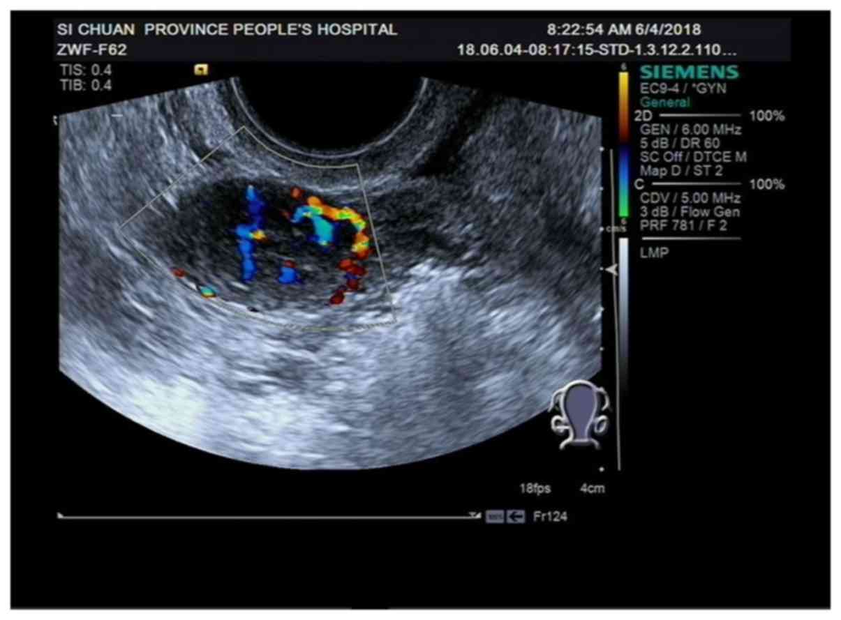
Using Color Doppler To Classify Ovarian Tumors Empowered Women S Health
Color Doppler Ultrasound Scan Of An Aneurysm Of The Abdominal Aorta Showing Circulating Lumen Of The Artery Orange Stock Photo Alamy
Comparison Of Image Features And Diagnostic Value Of Color Doppler Ultrasound And Two Dimensional Ultrasound In The Diagnosis Of Ovarian Sex Cord Stromal Tumors

The Color Doppler Ultrasound Image Of The Vertebral Artery A The Download Scientific Diagram

China Full Digital 3d 4d Color Doppler Ultrasound Machine Fdc8000 Photos Pictures Made In China Com
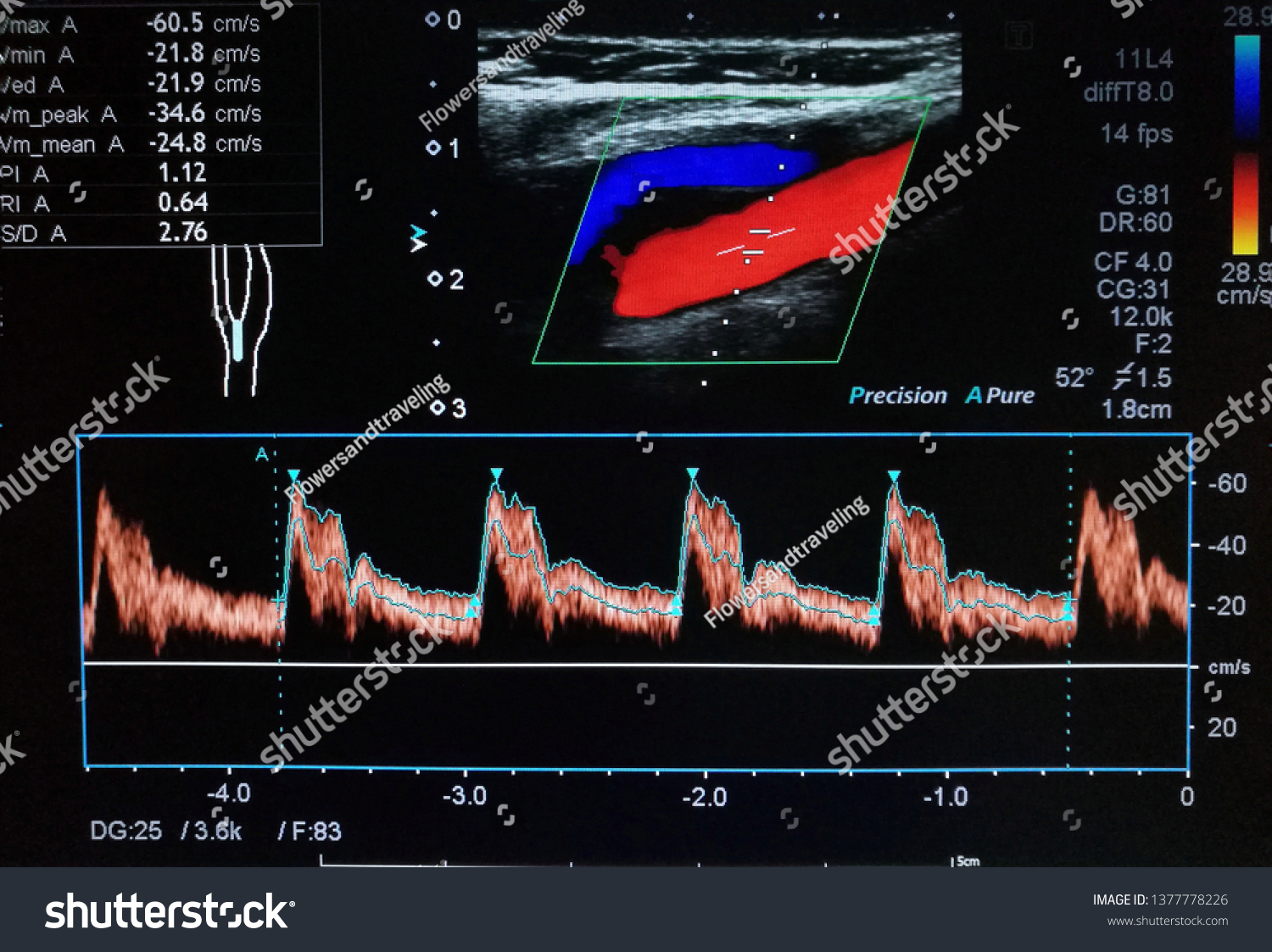
Color Doppler Ultrasound Imaging Normal Common Stock Photo Edit Now 1377778226
Color Doppler Ultrasound Images Shows Blood Flow Signals In The Wall Download Scientific Diagram
China Portable Color Doppler Ultrasound Scanner Photos Pictures Made In China Com
93+ Color Doppler Ultrasound Images. There are any 93+ Color Doppler Ultrasound Images in here.